Foreign Body Investigation & Microscopy
Foreign Body Investigation

The potential for damage to brand reputation is enormous, as is the potential for involvement in expensive litigation.
It is therefore essential that those involved in the food supply chain are able to respond quickly to every complaint with accurate information regarding the three main concerns:
What is it?
Where did it come from?
How did it get there?
Foreign Body Identification by Premier Analytical Services can help protect your brand integrity by quickly providing the information you require to determine the spurious, malicious or genuine nature of a complaint.
32 methods, accredited by UKAS to the ISO 17025 standard, assures analysis of the highest quality, thereby providing you with confidence in results you can trust.
Premier Analytical Services is one of the leading food testing centres in Europe and can place at your disposal a dedicated Microscopy team to solve your foreign body problems with decades of experience in foreign body identification.
Utilising “high-tech” facilities and non-destructive testing we offer a rapid and confidential service. Additional chemical and microbiological characterisation can also be carried out on foreign bodies, if required, using the additional facilities of the PAS laboratories. We can also arrange further independent expert identification and opinion from specialist consultants.
The results and expert interpretation are presented in a fully illustrated report with colour images and spectra, as appropriate. The analyses can be tailored to the customer’s requirements.
Full reports are routinely provided within 5 working days of sample receipt, in a PDF format via e-mail and a subsequent printed copy is returned by recorded delivery with the sample. A faster turnaround can be provided should your circumstances require an urgent response.
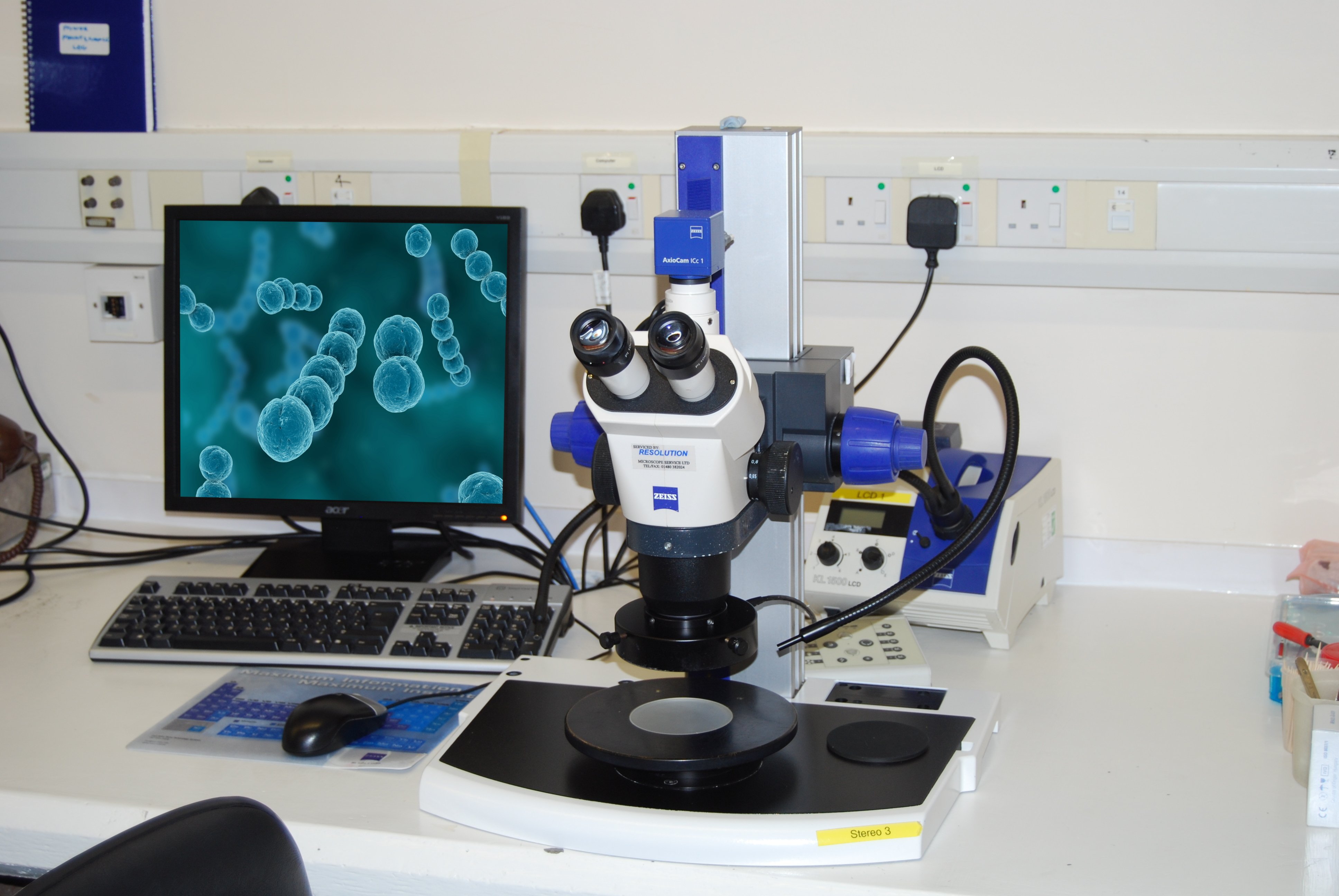
Optical Microscopy
Man-made and natural fibres, wood, rodent droppings, carbonised material, extraneous food materials, insects and hair are all examples of foreign bodies that can be identified by their morphological characteristics. Specific staining techniques can identify additional structural components. This provides information such as the presence, distribution and nature of any food material including starch, protein, fat/oil, cellular plant material and meat fibres.
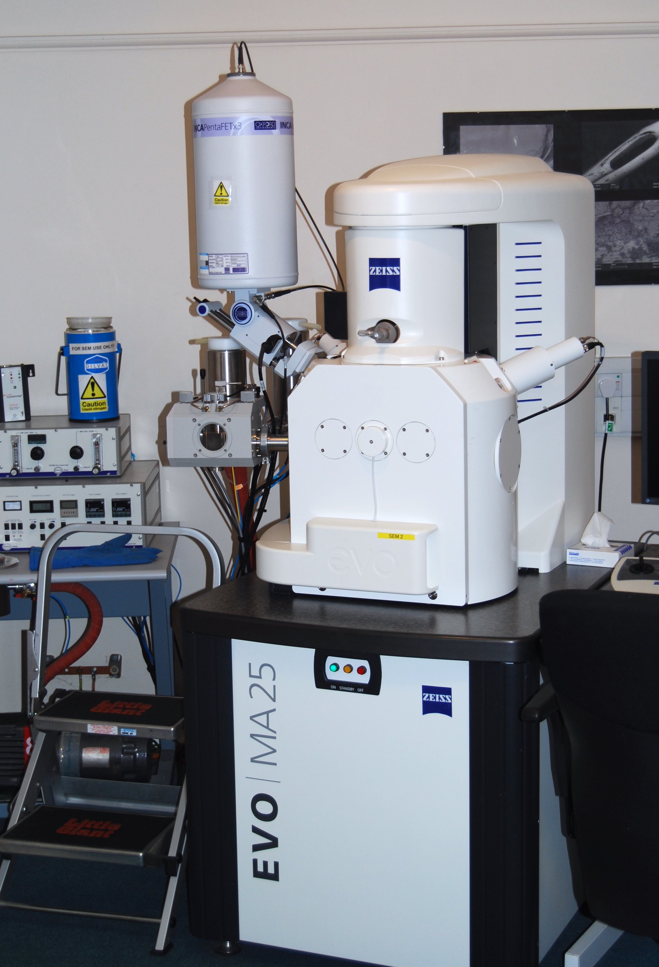
Scanning Electron Microscopy
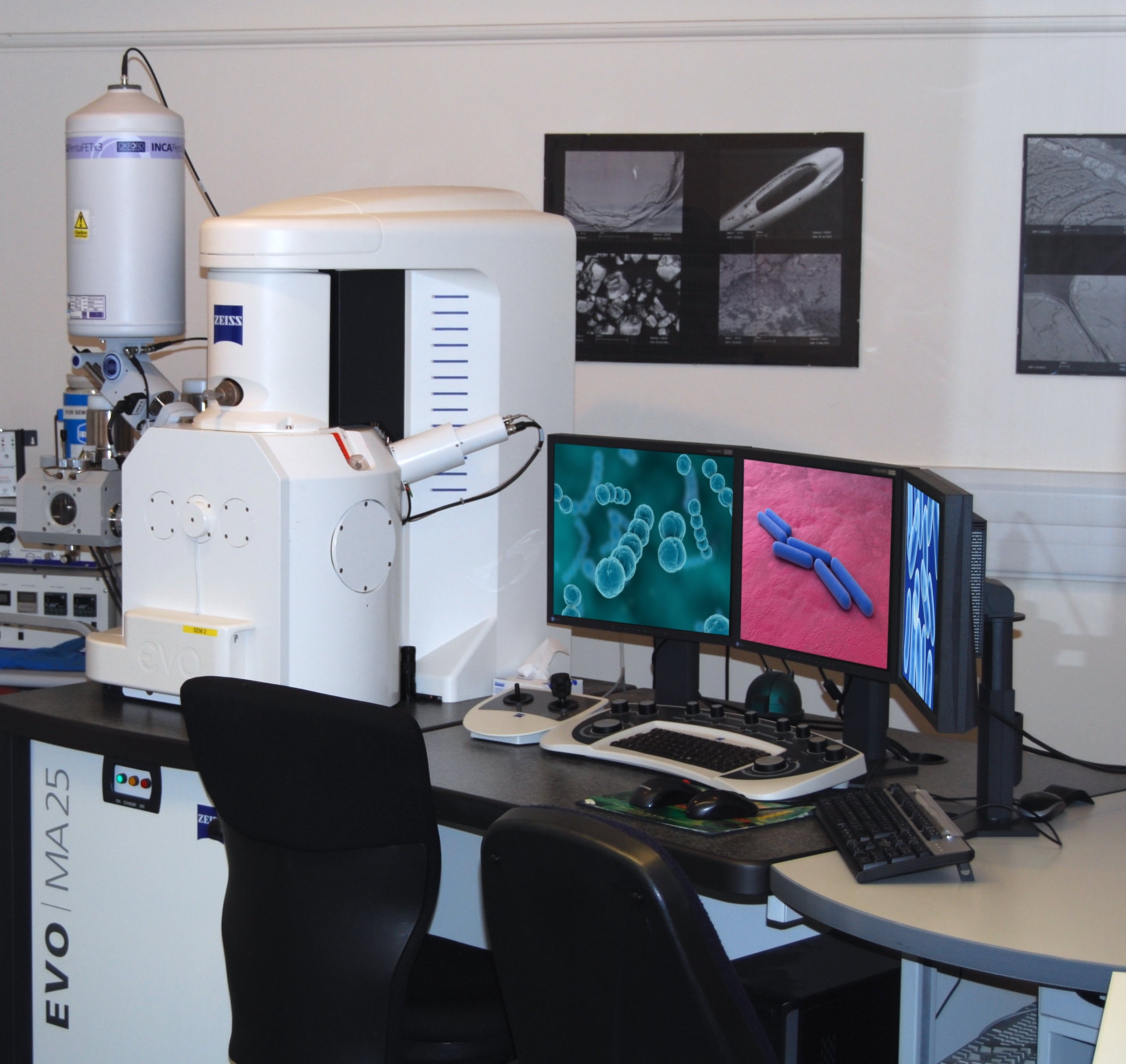
The scanning electron microscope gives topographical information concerning foreign objects including adhering deposits and their distribution. Areas of differing elemental composition can be rapidly identified indicating potential contact from metals and/or dental materials when dental damage is being claimed. This is useful in determining if any evidence of mechanical damage is present or if the sample has been bitten onto. A wide range of samples can be rapidly imaged and analysed without altering the evidence.
Energy Dispersive X-ray Analysis
Glass, metals and their alloys, dental amalgams and ceramics, tooth, bone and minerals are all examples of foreign bodies that can be identified by their elemental composition using this technique. This differentiates such materials as heat resistant and soda-lime glass, stainless steel types such as 200 and 300 series (austenitic) and 400 series (ferritic) stainless steel types and various alloys such as bronze and brass.
Fourier Transform Infra-Red Spectroscopy
Provides information about the chemical composition of materials. The spectra obtained can be searched against library spectra in order to identify a wide range of materials, including plastics, food materials, and man-made fibres.
Insect and Other Invertebrate Identification and Phosphatase Testing
Identification of insects and other invertebrates such as spiders, millipedes and slugs can be undertaken using key morphological features. Information on their habitats, habits and lifecycles can also be provided. Examination of the product packaging for damage may help further identify a point of contamination.
The active alkaline phosphatase enzyme is present in all living creatures and becomes denatured at temperatures of above approximately 70°C. Testing for the presence or absence of this enzyme is particularly useful with insect samples, where it can be determined if they have been heat processed. Combining this information with the analysis of any adhering deposits and evidence of damage to the packaging may be able to identify at what point contamination occurred. The test for the active alkaline phosphatase enzyme is destructive and requires certain sample conditions.
Alpha-Amylase (present in saliva) and Blood Testing
It may be important to determine if the sample has been in the mouth of the complainant. α-Amylase, a major component of saliva, can be rapidly identified on the sample by way of a spot test, which can confirm the presence or absence of α-amylase for such claims. The presence of blood can be similarly spot tested.
Reference Libraries and Databases
Our analyses and interpretation are supported by extensive in-house and external reference libraries and databases of materials, images, and spectra.
Customer Specific Databases
Databases of materials present within customer specific processes or factories can be compiled and used to facilitate the identification, or elimination, of possible foreign body sources.
Investigative Microscopy
Microscopical examination allows food structures to be visualised and thereby gives valuable information regarding the roles of the various raw ingredients and processing regimes on final product structure and attributes.
Premier Analytical’s investigative microscopy service can help you understand the effects of raw ingredients and processing on the appearance and texture of your products, enabling you to identify the cause of specific issues, improve product quality and develop new products with the desired attributes.
Premier Analytical Services’ Microscopy laboratory can help you to understand your products by providing expert interpretation of a range of complementary structural and compositional analyses:

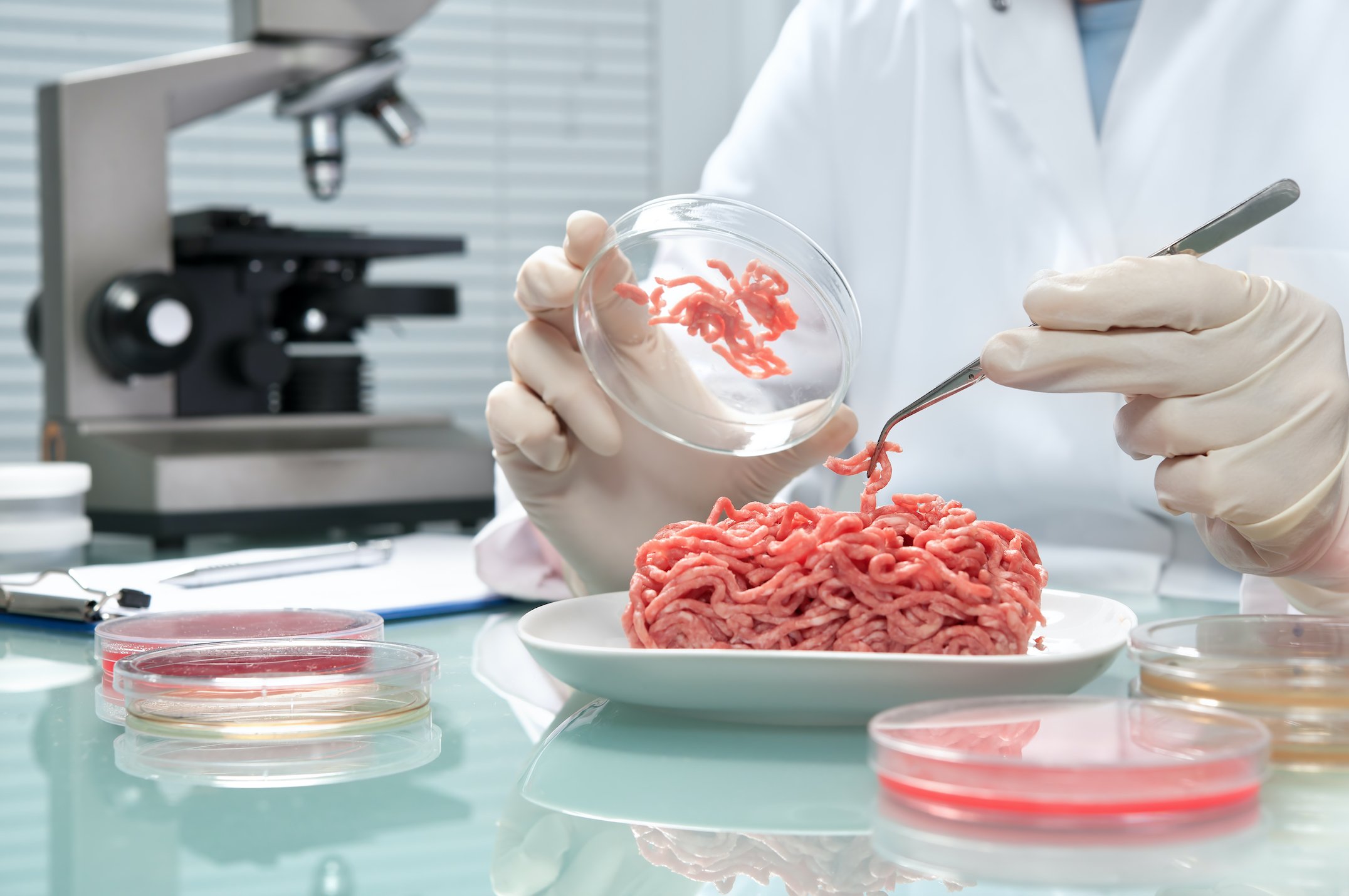
Optical microscopy – structures and spatial distributions can be elucidated for components such as fat/oil, protein, starch, cellular plant material, muscle fibres, gums and hydrocolloids.
Scanning electron microscopy – original and fracture surfaces of samples can be examined to give structural information relating to product appearance, aeration, density and texture.
Energy dispersive X-ray microanalysis – inorganic elemental composition can be determined to examine the distribution or dissolution of ingredients, such as salt, cocoa powder and fats.
Fourier transform infrared spectroscopy – determines the composition of polymer based packaging materials.
Image analysis – gives quantitative information on structural features, such as particle size / shape and product aeration / density.
The structures existing in raw ingredients affect how they process and their contribution to final product attributes. The selected processing regime can have a dramatic effect on the structure of the product components and therefore the texture/appearance of the final product.
Microscopy can aid in understanding raw ingredients and the structural changes they undergo through processing. Such information can then be used to optimise both processing efficiency and product quality. Microscopy can also be used to confirm some supplier’s ingredient claims.
A controlled heating stage allows heating processes to be mimicked on the optical microscope and changes to the sample structure monitored.
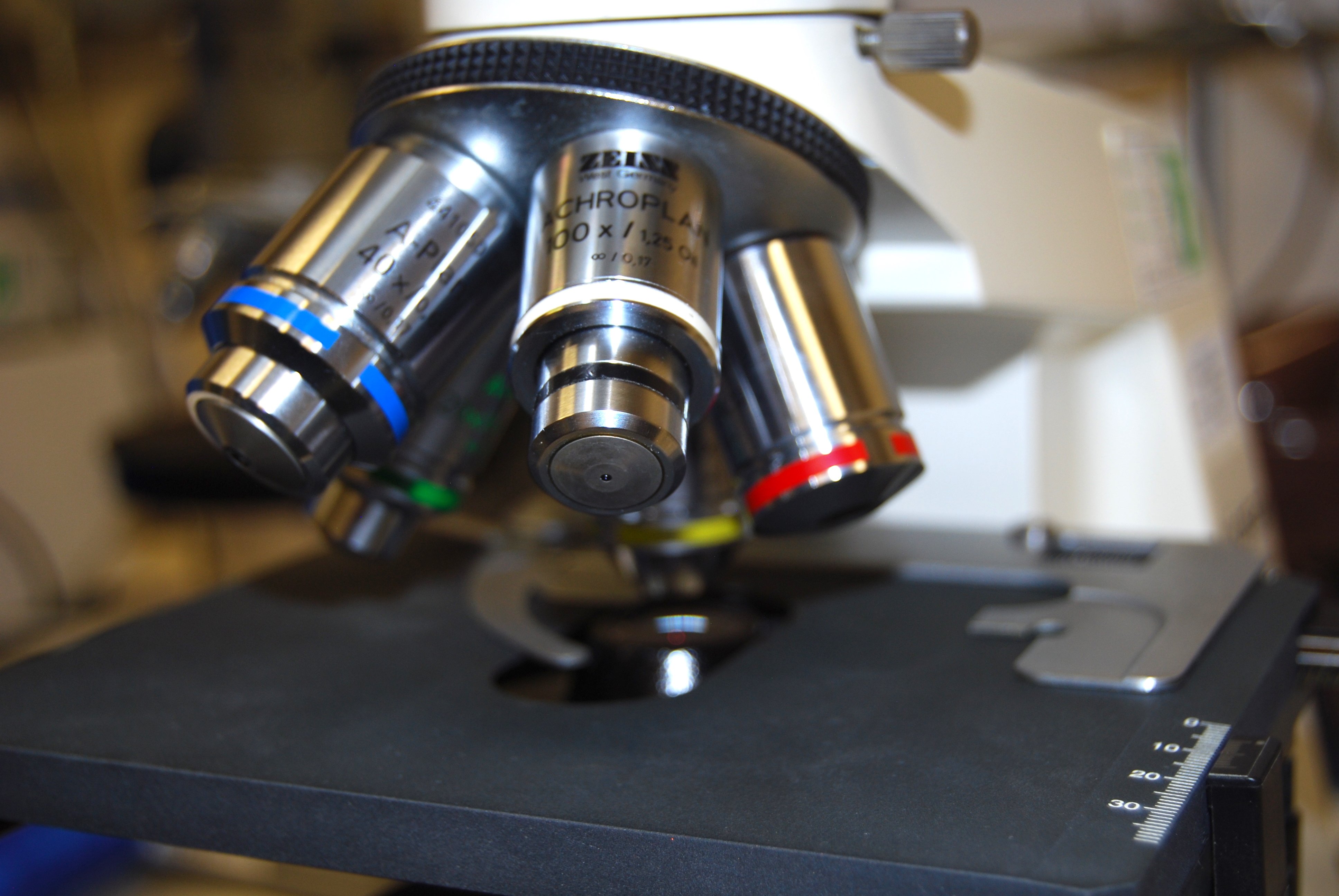
Final Product Quality – Appearance and Texture
An undesirable product appearance or texture, leading to poor product perception, can form during manufacture or develop over shelf-life. Achieving and maintaining the desired product attributes is therefore important. Identifying the cause of an unwanted characteristic is the first step to delaying or inhibiting its formation through reformulation, process changes or storage conditions.
Microscopical examination over the desired product shelf-life can allow the occurrence of undesired structural changes to be identified sooner, shortening the duration of unsuccessful shelf-life trials.
Equipment Assessment
The surface structures of processing equipment can affect product adhesion and release and therefore parameters such as process throughput. Examination of such equipment surfaces can help to explain their performance and be useful in the identification, or design, of better alternatives prior to their use / assessment on line.
Packaging Structure and Composition
Microscopy can be used to determine the structure of packaging materials, including cross-sectional and surface structures and seal formation. The additional use of Fourier transform infrared spectroscopy determines the composition of polymer type packaging materials. The results obtained can be searched against our libraries of reference spectra to determine the polymer type.
Foreign Body Investigation & Microscopy
Foreign Body Investigation

The potential for damage to brand reputation is enormous, as is the potential for involvement in expensive litigation.
It is therefore essential that those involved in the food supply chain are able to respond quickly to every complaint with accurate information regarding the three main concerns:
What is it?
Where did it come from?
How did it get there?
Foreign Body Identification by Premier Analytical Services can help protect your brand integrity by quickly providing the information you require to determine the spurious, malicious or genuine nature of a complaint.
32 methods, accredited by UKAS to the ISO 17025 standard, assures analysis of the highest quality, thereby providing you with confidence in results you can trust.
Premier Analytical Services is one of the leading food testing centres in Europe and can place at your disposal a dedicated Microscopy team to solve your foreign body problems with decades of experience in foreign body identification.
Utilising “high-tech” facilities and non-destructive testing we offer a rapid and confidential service. Additional chemical and microbiological characterisation can also be carried out on foreign bodies, if required, using the additional facilities of the PAS laboratories. We can also arrange further independent expert identification and opinion from specialist consultants.
The results and expert interpretation are presented in a fully illustrated report with colour images and spectra, as appropriate. The analyses can be tailored to the customer’s requirements.
Full reports are routinely provided within 5 working days of sample receipt, in a PDF format via e-mail and a subsequent printed copy is returned by recorded delivery with the sample. A faster turnaround can be provided should your circumstances require an urgent response.

Optical Microscopy
Man-made and natural fibres, wood, rodent droppings, carbonised material, extraneous food materials, insects and hair are all examples of foreign bodies that can be identified by their morphological characteristics. Specific staining techniques can identify additional structural components. This provides information such as the presence, distribution and nature of any food material including starch, protein, fat/oil, cellular plant material and meat fibres.
Scanning Electron Microscopy
The scanning electron microscope gives topographical information concerning foreign objects including adhering deposits and their distribution. Areas of differing elemental composition can be rapidly identified indicating potential contact from metals and/or dental materials when dental damage is being claimed. This is useful in determining if any evidence of mechanical damage is present or if the sample has been bitten onto. A wide range of samples can be rapidly imaged and analysed without altering the evidence.

Energy Dispersive X-ray Analysis
Glass, metals and their alloys, dental amalgams and ceramics, tooth, bone and minerals are all examples of foreign bodies that can be identified by their elemental composition using this technique. This differentiates such materials as heat resistant and soda-lime glass, stainless steel types such as 200 and 300 series (austenitic) and 400 series (ferritic) stainless steel types and various alloys such as bronze and brass.
Fourier Transform Infra-Red Spectroscopy
Provides information about the chemical composition of materials. The spectra obtained can be searched against library spectra in order to identify a wide range of materials, including plastics, food materials, and man-made fibres.
Insect and Other Invertebrate Identification and Phosphatase Testing
Identification of insects and other invertebrates such as spiders, millipedes and slugs can be undertaken using key morphological features. Information on their habitats, habits and lifecycles can also be provided. Examination of the product packaging for damage may help further identify a point of contamination.
The active alkaline phosphatase enzyme is present in all living creatures and becomes denatured at temperatures of above approximately 70°C. Testing for the presence or absence of this enzyme is particularly useful with insect samples, where it can be determined if they have been heat processed. Combining this information with the analysis of any adhering deposits and evidence of damage to the packaging may be able to identify at what point contamination occurred. The test for the active alkaline phosphatase enzyme is destructive and requires certain sample conditions.
Alpha-Amylase (present in saliva) and Blood Testing
It may be important to determine if the sample has been in the mouth of the complainant. α-Amylase, a major component of saliva, can be rapidly identified on the sample by way of a spot test, which can confirm the presence or absence of α-amylase for such claims. The presence of blood can be similarly spot tested.
Reference Libraries and Databases
Our analyses and interpretation are supported by extensive in-house and external reference libraries and databases of materials, images, and spectra.
Customer Specific Databases
Databases of materials present within customer specific processes or factories can be compiled and used to facilitate the identification, or elimination, of possible foreign body sources.
Investigative Microscopy

Microscopical examination allows food structures to be visualised and thereby gives valuable information regarding the roles of the various raw ingredients and processing regimes on final product structure and attributes.
Premier Analytical’s investigative microscopy service can help you understand the effects of raw ingredients and processing on the appearance and texture of your products, enabling you to identify the cause of specific issues, improve product quality and develop new products with the desired attributes.
Premier Analytical Services’ Microscopy laboratory can help you to understand your products by providing expert interpretation of a range of complementary structural and compositional analyses:

Optical microscopy – structures and spatial distributions can be elucidated for components such as fat/oil, protein, starch, cellular plant material, muscle fibres, gums and hydrocolloids.
Scanning electron microscopy – original and fracture surfaces of samples can be examined to give structural information relating to product appearance, aeration, density and texture.
Energy dispersive X-ray microanalysis – inorganic elemental composition can be determined to examine the distribution or dissolution of ingredients, such as salt, cocoa powder and fats.
Fourier transform infrared spectroscopy – determines the composition of polymer based packaging materials.
Image analysis – gives quantitative information on structural features, such as particle size / shape and product aeration / density.

The structures existing in raw ingredients affect how they process and their contribution to final product attributes. The selected processing regime can have a dramatic effect on the structure of the product components and therefore the texture/appearance of the final product.
Microscopy can aid in understanding raw ingredients and the structural changes they undergo through processing. Such information can then be used to optimise both processing efficiency and product quality. Microscopy can also be used to confirm some supplier’s ingredient claims.
A controlled heating stage allows heating processes to be mimicked on the optical microscope and changes to the sample structure monitored.

Final Product Quality – Appearance and Texture
An undesirable product appearance or texture, leading to poor product perception, can form during manufacture or develop over shelf-life. Achieving and maintaining the desired product attributes is therefore important. Identifying the cause of an unwanted characteristic is the first step to delaying or inhibiting its formation through reformulation, process changes or storage conditions.
Microscopical examination over the desired product shelf-life can allow the occurrence of undesired structural changes to be identified sooner, shortening the duration of unsuccessful shelf-life trials.
Equipment Assessment
The surface structures of processing equipment can affect product adhesion and release and therefore parameters such as process throughput. Examination of such equipment surfaces can help to explain their performance and be useful in the identification, or design, of better alternatives prior to their use / assessment on line.
Packaging Structure and Composition
Microscopy can be used to determine the structure of packaging materials, including cross-sectional and surface structures and seal formation. The additional use of Fourier transform infrared spectroscopy determines the composition of polymer type packaging materials. The results obtained can be searched against our libraries of reference spectra to determine the polymer type.
